The wide range of ultrasound machines available means that they can meet the differing needs of clinics, hospitals, and medical professionals. Below you’ll find the most common ultrasound machines available and what they can be used for.
Basic Ultrasound Machine
This is the most common type that typically produces two-dimensional images. The images are made using high-frequency sound waves that bounce off the internal structures of the body. The sound waves are received by a transducer which then creates the image.
Applications
- Listening to babies’ heartbeat during a sonogram
Pelvic Ultrasound Imaging
Pelvic ultrasound imaging is most commonly used for monitoring the health of a fetus or embryo during pregnancy. However, there are a number of other uses.
Applications
- Examining the uterus, bladder, ovaries, and prostate gland
- Diagnosing conditions such as abnormal bleeding, menstrual problems, pelvic pain, uterine fibroids, uterine and ovarian cancers
Transvaginal Ultrasound

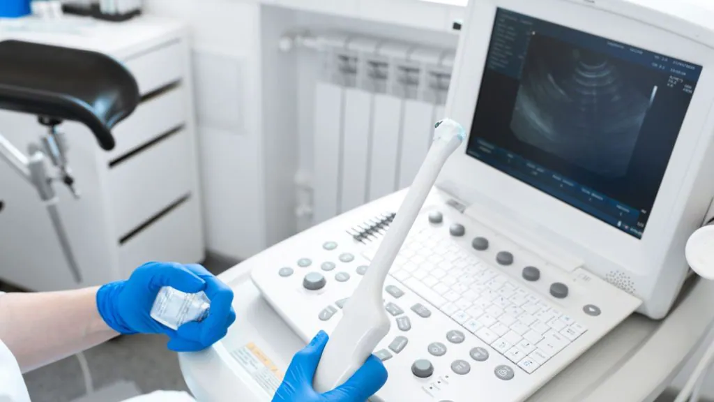
A transvaginal ultrasound scanner is used to see what’s going on inside a woman’s vagina. The examination takes place with the patient lying on their back with their feet in stirrups. A transducer is inserted into the vagina and moved around to capture images from different angles.
Uses:
- Detecting abnormalities
Transrectal Ultrasound
A transrectal imaging scanner is used to check inside the patient’s rectum. A transducer is inserted so that sound waves can travel to the prostate. The patient lies down for the examination on their left side.
Uses:
- Detecting abnormalities
Obstetric Imaging
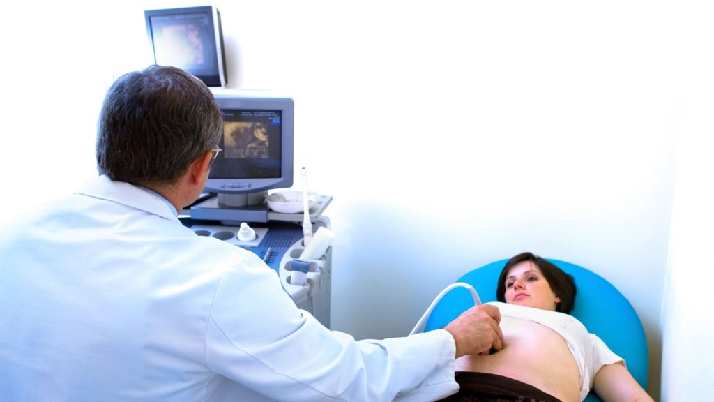
Obstetric imaging machines use sound waves to determine the condition of a pregnant woman and her fetus or embryo. The patient lies on their back or side, and they have to expose their lower abdominal area. The sonographer guides a transducer over the area. There is minimal discomfort.
Uses:
- Establishing the presence of a living fetus or embryo
- Estimating the pregnancy age
- Diagnosing congenital abnormalities
- Evaluating the position of the fetus
- Determining the location of the placenta
Breast Ultrasound Imaging
This type of ultrasound machine is specifically for scanning breast tissue. There is no radiation, so this is a preferred method for detecting breast cancer. The machine can detect the smallest abnormality. Doppler signals are also used to monitor blood flow.
Uses:
- Detecting early signs of breast cancer
- Ultrasound-guided biopsies for laboratory testing
- Monitoring blood flow
Abdominal Ultrasound Imaging
Abdominal ultrasound imaging machines are used to obtain images and examine internal organs and internal tissues to help diagnose a range of conditions or assess the damage caused by illness. It provides real-time ultrasound images of the liver, spleen, pancreas, bladder, gallbladder, and kidneys.
Uses:
- Guiding procedures such as needle biopsies
- Determining the source of abdominal pain
- Identifying the cause of an enlarged abdominal organ
- Doppler ultrasound models can evaluate blood flow, build-up of plaque, or congenital malformation
Kidney Imaging
A kidney or renal ultrasound machine uses sound waves to make images of the kidneys, bladder, and ureters.
Uses:
- Assessing the location, shape, and size of the kidneys and any related structures, such as the bladder and ureters
- Detecting tumors, cysts, obstructions, abscesses, infection, and fluid collection around and within the kidney
- Detecting calculi (stones) in kidneys and ureters
- Assisting with needle placement used in a biopsy of the kidneys or to drain fluid from an abscess or cyst
- Determining blood flow to the kidneys
- To evaluate a transplanted kidney
Uterus Imaging
This type of ultrasound machine looks at a woman’s uterus, tubes, ovaries, pelvic area, and cervix. The ultrasound probe is placed inside the vagina during the test.
Uses:
- Examining the shape, size, and position of the uterus and ovaries
- Measuring the length and thickness of the cervix
- Monitoring the blood flow through the various pelvic organs
- Identifying whether there are any changes to the shape of the bladder
- Diagnosing benign growths
- Endometriosis diagnosis
- Checking the heartbeat of a fetus